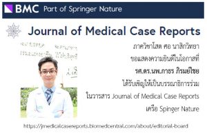
รศ.ดร.นพ.ภาธร ภิรมย์ไชย ได้รับเชิญเป็นบรรณาธิการร่วมในวารสาร Journal of Medical Case Reports

รศ.ดร.นพ.ภาธร ภิรมย์ไชย ได้รับเชิญเป็นบรรณาธิการร่วมในวารสาร Journal of Medical Case Reports ในเครือ Springer Nature

https://jmedicalcasereports.biomedcentral.com/about/editorial-board

รศ.ดร.นพ.ภาธร ภิรมย์ไชย ผ่าตัดไทรอยด์ผ่านกล้องชนิดซ่อนแผลใต้คางสำเร็จเป็นคนแรกของโลก

รศ.ดร.นพ.ภาธร ภิรมย์ไชย ภาควิชาโสต ศอ นาสิกวิทยา โรงพยาบาลศรีนครินทร์ คณะแพทยศาสตร์ มหาวิทยาลัยขอนแก่น ได้คิดค้นเทคนิคการผ่าตัดไทรอยด์ผ่านกล้องแบบใหม่ โดยซ่อนรอยแผลไว้ใต้คางและ/หรือหลังใบหู เรียกว่า post-auricular submental approach thyroidectomy หรือ PSA

เทคนิคใหม่นี้จะมีแผลขนาดประมาณ 1 เซนติเมตรที่ใต้คาง เซาะแผลใต้ผิวหนังน้อยลงกว่าเดิมครึ่งหนึ่ง ไม่มีแผลในช่องปาก จึงลดความเสี่ยงการติดเชื้อและการหายของแผลจะเร็วกว่า คนไข้สามารถรับประทานอาหารปกติตั้งแต่วันแรกหลังผ่าตัด อีกทั้งไม่มีแผลที่ทรวงอกหรือรักแร้ซึ่งไม่สวยงาม

อาจารย์ได้เริ่มผ่าตัดเทคนิคใหม่นี้ตั้งแต่ปี 2559 และได้รับการยอมรับตีพิมพ์ในวารสารของราชวิทยาลัยโสต ศอ นาสิกแพทย์แห่งประเทศอังกฤษ (the British Association of Otorhinolaryngology- Head & Neck Surgery) และสมาคมโสต ศอ นาสิกแพทย์เพื่อการวิจัยแห่งประเทศอังกฤษ (The Otorhinolaryngological Research Society)

สำหรับแพทย์ที่สนใจเทคนิคใหม่นี้ สามารถอ่านรายละเอียดได้ตาม link ด้านล่าง

https://onlinelibrary.wiley.com/doi/10.1111/coa.12791

#ผ่าตัดต่อมไทรอยด์ผ่านกล้อง #ผ่าตัดไทรอยด์แบบส่องกล้อง #โรงพยาบาลศรีนครินทร์ #มหาวิทยาลัยขอนแก่น #ขอนแก่น

ติดต่อโรงพยาบาลศรีนครินทร์

โทร. 043-366311, 043-366313

รศ.ดร.นพ.ภาธร ภิรมย์ไชย ได้รับเชิญเป็นบรรณาธิการวารสาร British Journal of Medicine and Medical Research

วารสาร British Journal of Medicine and Medical Research เป็นวารสารทางการแพทย์นานาชาติ ที่เปิดให้อ่านได้ฟรีไม่ต้องสมัครสมาชิก วารสารได้รับการจัดเก็บไว้ในฐานข้อมูล US National Library of Medicine (NLM) Catalog, ISI Web of Science citations, Index Medicus, ProQuest, EBSCOhost, DOAJ และอื่น ๆ

บรรณาธิการของวารสารนี้มาจากทั้งสหรัฐอเมริกา ยุโรปและเอเชีย โดยผศ.ดร.นพ.ภาธร ภิรมย์ไชย เป็นบรรณาธิการที่เป็นคนไทยเพียงผู้เดียว ท่านสามารถเข้าดูรายชื่อบรรณาธิการได้ตาม link ด้านล่าง

http://www.sciencedomain.org/journal/12/editorial-board-members

Correlations of External Landmarks with Internal Structures of the Temporal Bone

Piromchai P, Wijewickrema S, Smeds H, Kennedy G, O’Leary S.

Otol Neurotol. 2015 Jul 17.

Abstract:

The internal anatomy of a temporal bone could be inferred from external landmarks.

BACKGROUND:

Mastoid surgery is an important skill that ENT surgeons need to acquire. Surgeons commonly use CT scans as a guide to understanding anatomical variations before surgery. Conversely, in cases where CT scans are not available, or in the temporal bone laboratory where residents are usually not provided with CT scans, it would be beneficial if the internal anatomy of a temporal bone could be inferred from external landmarks.

METHODS:

We explored correlations between internal anatomical variations and metrics established to quantify the position of external landmarks that are commonly exposed in the operating room, or the temporal bone laboratory, before commencement of drilling. Mathematical models were developed to predict internal anatomy based on external structures.

RESULTS:

From an operating room view, the distances between the following external landmarks were observed to have statistically significant correlations with the internal anatomy of a temporal bone: temporal line, external auditory canal, mastoid tip, occipitomastoid suture, and Henle’s spine. These structures can be used to infer a low lying dura mater (p = 0.002), an anteriorly located sigmoid sinus (p = 0.006), and a more lateral course of the facial nerve (p < 0.001). In the temporal bone laboratory view, the mastoid tegmen and sigmoid sinus were also regarded as external landmarks. The distances between these two landmarks and the operating view external structures were able to further infer the laterality of the facial nerve (p < 0.001) and a sclerotic mastoid (p < 0.001). Two nonlinear models were developed that predicted the distances between the following internal structures with a high level of accuracy: the distance from the sigmoid sinus to the posterior external auditory canal (p < 0.001) and the diameter of the round window niche (p < 0.001).

CONCLUSION:

The prospect of encountering some of the more technically challenging anatomical variants encountered in temporal bone dissection can be inferred from the distance between external landmarks found on the temporal bone. These relationships could be used as a guideline to predict challenges during drilling and choosing appropriate temporal bones for dissection.

http://dx.doi.org/10.1097/MAO.0000000000000824

Impact of treatment time on the survival of patients suffering from invasive fungal rhinosinusitis.

Abstract

BACKGROUND:

Invasive fungal rhinosinusitis is an uncommon disease with high mortality rates. There is currently no consensus on the best treatment timing. We studied the impact of the treatment timing on the survival of patients experiencing invasive fungal rhinosinusitis.

METHODS:

We conducted a retrospective study of patients suffering from invasive fungal rhinosinusitis. The duration of symptoms, clinical presentations, clinical signs, diagnoses, treatments, and outcomes were collected.

RESULTS:

It was observed that more than 70% of the mortalities occurred within the subgroup of patients who exhibited symptoms of the disease within 14 days before admission. After adjusting for the confounders, the time taken to treat the patients was the most statistically significant predictor for mortality (P = 0.045). We found no significant relationships between mortality and its significant covariates, which included the underlying diseases (P = 0.91) or complications (P = 0.55).

CONCLUSIONS:

Our study demonstrates that the time taken to treat the patients is an important determinant for the survival of patients who are afflicted with invasive fungal rhinosinusitis. The appropriate treatments should be administered within 14 days from the time the symptoms begin to manifest.

http://www.ncbi.nlm.nih.gov/pubmed/25288891

The construct validity and reliability of an assessment tool for competency in cochlear implant surgery.

Abstract

INTRODUCTION:

We introduce a rating tool that objectively evaluates the skills of surgical trainees performing cochlear implant surgery.

METHODS:

Seven residents and seven experts performed cochlear implant surgery sessions from mastoidectomy to cochleostomy on a standardized virtual reality temporal bone. A total of twenty-eight assessment videos were recorded and two consultant otolaryngologists evaluated the performance of each participant using these videos.

RESULTS:

Interrater reliability was calculated using the intraclass correlation coefficient for both the global and checklist components of the assessment instrument. The overall agreement was high. The construct validity of this instrument was strongly supported by the significantly higher scores in the expert group for both components.

CONCLUSION:

Our results indicate that the proposed assessment tool for cochlear implant surgery is reliable, accurate, and easy to use. This instrument can thus be used to provide objective feedback on overall and task-specific competency in cochlear implantation.

http://www.ncbi.nlm.nih.gov/pubmed/25114897

Virtual Reality Surgical Training in Ear, Nose and Throat Surgery

ABSTRACT

Virtual reality has been introduced to the medical field and it is now used in medical education as an alternative high fidelity simulator. The major components to successful learning that has contributed to the efficacy of the virtual reality system are the ability to provide repetitive practice under controlled environment, self-directed learning and proved construct validity. This review evaluated the validity and the efficacy of current virtual reality systems in ear, nose and throat surgery.

http://www.scirp.org/journal/PaperInformation.aspx?PaperID=45833

Invasive Fungal Rhinosinusitis versus Bacterial Rhinosinusitis with Orbital Complications: A Case-Control Study.

Abstract

Background. Invasive fungal rhinosinusitis with orbital complications (IFSwOC) is a life-threatening condition. The incidence of mortality has been reported to be up to 80 percent. This study was conducted to determine the risk factors, presentations, clinical, and imaging findings that could help to manage this condition promptly. Methods. We conducted a case-control study of 100 patients suffering from rhinosinusitis with orbital complications. The risk factors, clinical presentations, radiological findings, medical and surgical managements, durations of hospital stay, and mortality rate data were collected. Results. Sixty-five patients were diagnosed with IFSwOC, while the other thirty-five patients composed the control group. The most important risk factor for IFSwOC was diabetes mellitus. Visual loss and diplopia were the significant symptom predictors. The significant clinical predictors were nasal crust, oculomotor nerve, and optic nerve involvement. The CT findings of IFSwOC were sinus wall erosion and hyperdensity lesions. The mortality rate was 25.71 percent in the IFSwOC group and 3.17 percent in the control group. Conclusions. Invasive fungal rhinosinusitis with orbital complications is symptomatic of a high mortality rate. The awareness of a patient’s risk factors, the presenting symptoms, signs of fungal invasion, and aggressive management will determine the success of any treatment procedures.

http://www.ncbi.nlm.nih.gov/pubmed/24298218

Chronic rhinosinusitis and emerging treatment options

This review describes the epidemiology and various treatments in chronic rhinosinusitis (CRS) with nasal polyps (CRSwNP) and CRS without nasal polyps (CRSsNP). Evidence for short-term use of systemic corticosteroids has been shown to be favorable in CRSwNP, but still limited in CRSsNP. Topical corticosteroids improve symptom scores in both CRS subgroups. The role of microbes in CRS is still controversial. Culture-directed antibiotics are recommended for CRSsNP with exacerbation. Long-term use of low dosage antibiotics is recommended for CRSsNP for their anti-inflammatory effects. Other emerging treatment options are also discussed.

http://www.dovepress.com/chronic-rhinosinusitis-and-emerging-treatment-options-peer-reviewed-article-IJGM

Acute Versus Chronic Invasive Fungal Rhinosinusitis: A Case-Control Study

Authors: Patorn Piromchai and Sanguansak Thanaviratananich

Introduction: Invasive fungal rhinosinusitis is a challenging condition that can be found mostly in immunocompromised patients. Failure to diagnose and treat this entity promptly usually results in rapid progression and death. The purpose of this study was to determine clinical presentation, complication and morbidity in patients with acute versus chronic invasive fungal rhinosinusitis.

Setting and design: Case-control study at Srinagarind Hospital, Khon Kaen University between January 1998 and May 2008.

Methods: The patient’s data with the diagnosis of invasive fungal rhinosinusitis was included. Demographic data, underlying diseases, presenting symptoms, histologic sinonasal tissue evaluations, sinonasal tissue cultures, CT scan findings, surgical interventions, morbidity, and mortality were collected.

Results: Sixty-five patients were diagnosed as invasive fungal rhinosinusitis between January 1998 and May 2008. The data of six patients were unable to obtain. Fifty-nine patients were included in this study. Patients with immunocompromised status have significant greater risk for acute than chronic IFS, OR = 6.5 (P = 0.004). Patients with mucosal necrosis have the significant higher risk for acute IFS, OR = 5.5 (P = 0.01). There was no significant difference in orbital complications proportion between acute and chronic invasive fungal rhinosinusitis, OR = 2.42 (P = 0.15). Sinus wall erosion have found significantly in chronic IFS group, OR = 0.24 (P = 0.02). The average hospital stayed was 30.58 ± 26.43 days with no difference between groups (P = 0.50). Fourteen patients in acute IFS group were dead (31.11%) while all patients in chronic IFS group were survived.

Conclusions: Invasive fungal rhinosinusitis continues to present a challenge to the otolaryngologist. Acute IFS was found most commonly in immunocompromised patients. The most consistent finding of acute IFS was mucosal necrosis and black crust/debris. The CT finding of sinus wall erosion may help in diagnosis of chronic IFS.

http://www.la-press.com/acute-versus-chronic-invasive-fungal-rhinosinusitis-a-case-control-stu-article-a3233